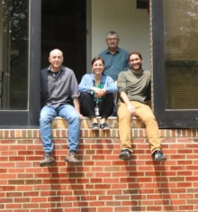
Dr. Natalia Gudino received an Electrical Engineering degree with honors from the National University of Rosario, Argentina in 2003. She pursued her graduate training at Case Western Reserve University (CWRU). First, as a Fulbright Scholar, she developed a steerable catheter controlled by the MRI system at Dr. Mark Griswold’s lab.
After earning her M.Sc. (2008) in Biomedical Engineering, she started her Ph.D. research working on the development of on-coil RF amplification to implement parallel transmission (pTx) in MRI. In a joint collaboration with Dr. Robert Lederman and Dr. Robert Balaban (NHLBI), she explored the impact of this technology on improving RF safety in an interventional MRI setup. After receiving her Ph.D. in 2013, she joined, Dr. Jeff Duyn’s lab at LFMI, NINDS, to develop new on-coil RF amplifiers that can be utilized in ultra-high field MRI (≥7T), and a novel electronic interface that allows control of the new technology by a broad range of commercial systems.
She became a Staff Scientist at the LFMI in 2018 and leads the MRI Engeineering Team (MRIEngT). Dr. Gudino is an active member of the International Society of Magnetic Resonance in Medicine and a recipient of the 2017 outstanding teacher award for lecturing RF engineering. As a Staff Scientist and Leader of the MRI Engineering Team, her research mainly focuses on new technologies for RF transmission and signal reception to improve brain imaging at ultra-high field.
MRI Engineering Team (MRIEngT) Overview
The overall goal of the MRI Engineering Team (MRIEngT) is to generate new hardware technologies to develop the next generation of MRI systems, especially for high field MRI. The MRIEngT is part of the Laboratory of Functional and Molecular Imaging (LFMI) and it is located at the NMR Center, with access to a variety of MRI systems, from low to ultra-high magnetic field scanners (7 T and above). The current focus is on getting one of the first human 11.7 T MRI operational. In addition, the team works on several projects to optimize existing MRI applications across the magnetic field spectrum for human and animal imaging (from 64 mT to 14 T). This work demands the customized design of innovative hardware and software.
The MRIEngT is particularly interested on the advancement of the MRI transmit and receive architectures through the design and implementation of new, low-cost, power efficient and compact electronics. This new hardware is expected to improve imaging of the brain at ultra-high field MRI helping the LFMI in its mission to further understand brain function. On this effort, engineering methods and technologies generated by the team can also improve imaging beyond the brain.
Current Research
Parallel Tx hardware for Ultra High Field MRI
A major challenge at ultra-high field MRI is the compensation of the transmit field (B1) inhomogeneities that result from the interaction of the shorter wavelength excitation with the tissue. These inhomogeneities lead to spatially varying contrast, and more focal energy depositions that may impose safety limitations on some applications.
Parallel transmission (pTx) allows independent control of the amplitude and phase of the multiple linearly polarized fields generated in a transmit (Tx) coil array. This flexibility is used to shim the transmit field as well as to control the associated transmission electrical field responsible for tissue heating.
Most pTx hardware today are built with the 50 Ohm coaxial architecture used in single channel transmitters. However, the implementation of pTx systems with higher channel count at ultra-high field MRI demands the development of dedicated Tx hardware. Optically controlled and monitored on-coil current-source RF power amplifiers (RFPAs) allow the implementation of a pTx chain without cable losses and coupling and minimal load sensitivity at a lower implementation cost. This technology allows direct sensing of coil current for safety monitoring and feedback. The MRIEngT is further developing this technology and its control for operation at higher magnetic fields and frequencies.
- Setera B, Christou A and Gudino N. Monitoring Current of GaN HEMT at Ultra-High Magnetic Fields. IEEE 10th Workshop on Wide Bandgap Power Devices & Applications (WiPDA) 2023. Page 1-5
- Gudino N, de Zwart JA, Duyn JH. Eight-channel parallel transmit-receive system for 7 T MRI with optically controlled and monitored on-coil current-mode RF amplifiers. Magn Reson Med. 2020 Dec;84(6):3494-3501.
- Gudino N, de Zwart JA, Duan QI, et al. Optically controlled on-coil amplifier with RF monitoring feedback. Magn Reson Med. 2018;79:2833-2841.
- Gudino N, Duan QI, de Zwart JA, et al. Optically controlled switch-mode current-source amplifiers for on-coil implementation in high-field parallel transmission. Magn Reson Med. 2016;76:340-349.
Parallel Rx hardware for Ultra High Field MRI
Rx coil arrays are essential hardware to achieve maximum SNR at ultra-high field MRI. The MRIEngT is developing 11.7 T Rx head arrays to be combined with different Tx hardware. To simplify the implementation and boost performance of high-density Rx arrays, the team is also working on the optimization of in-house built low-noise amplifier and novel detuning electronics.
- Dodd SJ, Murphy-Boesch J, Merkle H, Silva A, Koretsky AP. A 12-element Receive Coil Array for the Rat Brain at 11.7 T. In: Proc. Intl. Soc. Mag. Reson. Med. 20; 2012:437.
Novel hardware for Multinuclear MRI
Multinuclear MRI and MRS allows the study of brain metabolism non-invasively and the detection of biomarkers in diseases such as Alzheimer and cancer. The MRIEngT is developing new multinuclear coils and hardware that will add flexibility to the MRI system for routine implementation of studies that involve detection of multiple nuclei.
- Gudino N, Adaptable Dual-Tuned Optically Controlled On-Coil RF Power Amplifier for MRI. IEEE Trans Biomed Circuits Syst. 2024 Jun 5. Online ahead of print.
- Gudino N, Dodd S, Li S et al. Dual-Tuned Optically Controlled On-Coil Switch-Mode Amplifier. Proceedings of the 28th Annual meeting, ISMRM, Virtual 2020. (Oral Presentation, Abstract 0751)
- Gudino N. Adaptable Dual-Tuned Optically Controlled On-Coil Amplifier for High-Field MRI Systems. US Patent Application 20230243905A1
RF safety assessment of in-house built hardware
In MRI the RF hardware generates conservative and magnetically induced electrical fields. These fields are the source of tissue heating, and therefore their minimization is required to ensure patient safety. A commonly used safety parameter is the Specific Absorption Rate (SAR) in watts per kilogram which indicates how much power is absorbed by the tissue. SAR can be predicted by performing electromagnetic field simulations using a model of the RF hardware and tissue. The MRIEngT dedicates efforts to modeling, simulating and designing experimental validation methods to assess the safety of in-house built hardware.
- Gudino N, de Zwart JA, Dodd S, et al. Model validation of a 500 MHz Inductive Resonator for 11.7 T Brain MR. Proceedings of the 30th Annual meeting, ISMRM, London, UK 2022. (Poster, Abstract 2097)
- Murphy-Boesch J, de Zwart JA, van Gelderen P, et al. 500 MHz Inductive Birdcage RF Coil for Brain MRI: Design, Implementation and Validation. IEEE Transactions on Biomedical Engineering, doi: 10.1109/TBME.2025.3529725.
Development of new hardware for various MRI applications
Arterial spin labeling (ASL) allows imaging perfusion in the brain. Continuous ASL, in the brain has been demonstrated using a separate labeling surface coil driven by a conventional remote power amplifier. This setup presents potential crosstalk between the long coaxial connection to the labeling coil and the imaging RF hardware, coaxial losses and sensitivity to patient loading. This can be improved by replacing the remote voltage-mode RF amplifier (amplifier located outside MRI room) by an optically controlled on-coil current-source switch-mode RF amplifier. The MRIEngT aims to develop a dedicated on-coil amplifier and control to perform ASL in the brain at ultra-high field MRI.
- Saïb G, Huang S, Long E et al. Continuous Arterial Spin Labeling with On-Coil Amplification at 7T. Proceedings of the 30th Annual meeting, ISMRM, London, UK 2022. (Poster, Abstract 0646)
Team Members
Selected Publications
Gudino N. Adaptable Dual-Tuned Optically Controlled On-Coil RF Power Amplifier for MRI. IEEE Trans Biomed Circuits Syst. 2024 Jun 5. Online ahead of print.
Gudino N, de Zwart JA, Duyn JH. Eight-channel parallel transmit-receive system for 7 T MRI with optically controlled and monitored on-coil current-mode RF amplifiers. Magn Reson Med. 2020 Dec;84(6):3494-3501.
Gudino N, de Zwart JA, Duan Q, Dodd SJ, Murphy-Boesch J, van Gelderen P, Duyn JH. Optically controlled on-coil amplifier with RF monitoring feedback. Magn Reson Med. 2018 May;79(5):2833-2841.
Qian C, Duan Q, Dodd SJ, Koretsky AP, Murphy-Boesch J. Sensitivity Enhancement of an Inductively Coupled Local Detector Using a HEMT-Based Current Amplifier. Magn Reson Med. 2016 Jun;75(6):2573-8.
Duan Q, Nair G, Gudino N, de Zwart JA, van Gelderen P, Murphy-Boesch J, Reich DS, Duyn JH, Merkle H. A 7T spine array based on electric dipole transmitters. Magn Reson Med. 2015 Oct;74(4):1189-97.
Duan Q, Duyn JH, Gudino N, de Zwart JA, van Gelderen P, Sodickson DK, Brown R. Characterization of a dielectric phantom for high-field magnetic resonance imaging applications. Med Phys. 2014 Oct;41(10):102303.
Merkle H, Murphy-Boesch J, van Gelderen P, Wang S, Li TQ, Koretsky AP, Duyn JH. Transmit B1-field correction at 7 T using actively tuned coupled inner elements. Magn Reson Med. 2011 Sep;66(3):901-10.

