
Know Stroke
Stroke causes severe life-changing
disruption.
Together, we can
disrupt stroke.
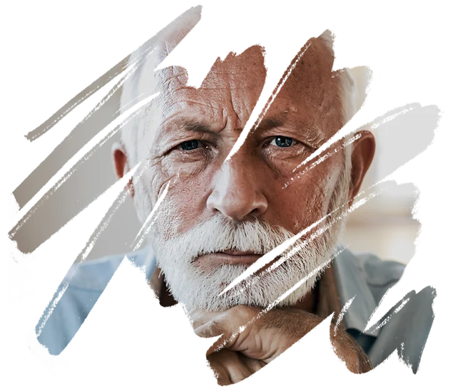
Stroke, a leading cause of death, can take away your ability to talk, walk, and think clearly. Stroke can happen to anyone—even if you’re young. Let’s disrupt this disruptor with prevention, treatment, and research.
Know The Symptoms
Act F.A.S.T.
to Save Lives
-
FFace
DroopingDoes one side of the face droop when smiling?
-
AArm
WeaknessWhen arms are raised, does one drift down?
-
SSpeech
DifficultyIs speech slurred or strange?
-
TTime to
Call 911Do not drive — call an ambulance immediately.
Every second counts.
Stroke is a medical emergency. About 80% are ischemic strokes, which cut off blood to the brain. The rest are hemorrhagic strokes, caused by bleeding in or around the brain. The longer blood flow is cut off, the greater the damage. Getting to a hospital quickly saves lives and increases the chances for successful recovery.
Explore
Know Stroke Resources
-
Prevention
Learn the signs of stroke and what to do if you think someone is having a stroke.
-
Signs &
SymptomsKnow all the sudden symptoms of stroke and act F.A.S.T. to recognize stroke and get immediate treatment.
-
Assess &
TreatExplore assessment and treatment tools, research, and resources, including the NIH Stroke Scale.
-
Recovery
Stay on top of the latest stroke related research conducted and funded by NINDS, NIH, and major medical institutions.
-
Research
Stay on top of the latest stroke related research conducted and funded by NINDS, NIH, and major medical institutions.
-
Outreach
Share Know Stroke campaign materials to raise awareness to prevent and treat stroke in your community.
-
Stroke
ScaleLearn about and access the widely used NIH Stroke Scale, which helps doctors assess the severity of a stroke.
-
Clinical
TrialsLearn about the steps and safety measures in clinical trials and how to participate.
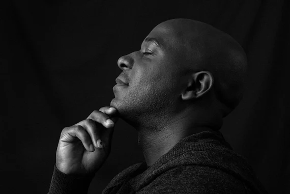
Special Feature
Nobody can reduce your risk like you.
Everyone is at risk for stroke and dementia. But if you’re a Black man 28-45, you have a higher risk. Many years before stroke or dementia happen, uncontrolled high blood pressure narrows your arteries, decreasing blood to your brain. Take charge of your health!